Endoscopic Mucosal Resection and Endoscopic Submucosal Dissection
Endoscopic Mucosal Resection
Endoscopic mucosal resection (EMR) is a technique used for the staging and treatment of superficial neoplasms of the gastrointestinal tract. The technique was first developed in Japan for the treatment of early gastric cancer and has since spread in use throughout the world for various indications, including dysplastic Barrett mucosa and sessile colonic neoplasms. The utility of EMR rests in its ability to do the following:
- Provide accurate histological staging of superficial GI neoplasms
- Provide a minimally invasive technique for removal of superficial malignancies.
Several variations of EMR are currently used, including injection-assisted, cap-assisted, and ligation-assisted techniques. All adhere to the basic principles of identification and demarcation of the lesion, submucosal injection to lift the lesion, and endoscopic snare resection. Due to its overall safety and efficacy in appropriately selected patient populations, EMR has become firmly integrated in the diagnostic and treatment algorithms of superficial GI malignancies.
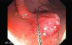
Major advances in the development of endoscopic devices and techniques over the past fifteen years have introduced endoscopic mucosal resection (EMR) and endoscopic submucosal dissection (ESD) as standard of care for the safe and effective removal and/or definitive staging of mucosal lesions of the esophagus, stomach, duodenum and colon, often eliminating major surgery as first-line management.
With EMR, normal saline or hydroxy propyl methylcellulose, dilute epinephrine and methylene blue are injected into key areas of the submucosal space beneath the tumor, strategically positioning the tumor and separating it from the bowel wall; the tumor can then be resected with less risk of thermal or mechanical damage to the muscularis propria.
More On EMR
EMR is an endoscopic technique developed for removal of sessile or flat neoplasms confined to the superficial layers (mucosa and submucosa) of the GI tract. EMR is
typically used for removal of lesions smaller than 2 cm or piecemeal removal of larger lesions. Most commonly used techniques can be subdivided as injection-, cap, and ligation-assisted EMR. Before the start of any EMR technique, it may be helpful to mark the margins of a tar-geted lesion with superficial cautery marks. Injection-assisted EMR, also often called ‘‘saline-assis-ted’’ polypectomy, is frequently used for large flat colon polyps. This technique was introduced in 1955 for rigid sig-moidoscopy and then in 1973 for flexible colonoscopy. The procedure starts with injection of a solution into the submucosal space under the lesion, creating a ‘‘safety cush-ion.’’ The cushion lifts the lesion to facilitate its removal and minimizes mechanical or electrocautery damage to the deep layers of the GI tract wall. Injection-assisted EMR can be further subdivided into the ‘‘inject-and-cut’’ technique (using an electrocautery snare through a sin-gle-channel endoscope) or the ‘‘inject-lift-and-cut’’ tech-nique (using a grasping forceps to lift the lesion and an electrocautery snare through separate channels of a dou-ble-channel endoscope).
As a variation of the latter tech-nique, EMR of large gastric lesions may be assisted by countertraction of the lesion with a grasping forceps placed through a percutaneous endoscopic gastrostomy tract. To facilitate the basic processes of injection and snare excision with instrument, a device combining these functions has recently been developed.
Endoscopic Submucosal Dissection
ESD has been developed for en bloc removal of large (usually more than 2 cm), flat GI tract lesions. The pro-cedure is usually done in several steps. First, the margins of the lesion are marked by electrocautery, and submucosal injection is used to lift the lesion. Then, a circumferential incision into the submucosa is performed around the lesion with specialized endoscopic electrocautery knives. Finally, the lesion is dissected from underlying deep layers of GI tract wall with the electrocautery knife and removed. After removal, the en bloc pathologic specimen should be mounted and oriented to facilitate histologic examination. Multiple cutting devices and accessories have been de-veloped specifically for ESD. These devices are not yet commercially available in the United States.
Polypectomy
Overview
Polypectomy is the medical term for removing polyps. Colonic polyps are abnormal like growths that protrude into the lining of the bowel. Small polyps can be removed by an instrument called a biopsy forceps, which snips off small pieces of tissue. Larger polyps are usually removed by putting a noose, or snare, around the polyp base and burning through the tissue with electric cautery. Neither of these procedures is painful,and you will usually not be aware that they are being done. Rarely will a polyp be too large to remove by colonoscopy and require surgery for removal.
Polypectomy is very safe, but all procedures entail some risks, which you should discuss with your doctor. The most common complications of polypectomy include bleeding and perforation (creating a hole in the colon). Fortunately, although these are the most common complications of polypectomy, they are still infrequent. Bleeding can usually be controlled by colonoscopy, during which the bleeding site is cauterized, although surgery is sometimes required. Surgery is usually required for perforation. Other complications have been described but occur much less frequently.
Polyps
Colonic polyps are abnormal like growths that protrude into the lining of the bowel. There are two types of polyps: pedunculated and sessile. Pedunculated polyps appear almost mushroom-like. That is, the polyp has a stalk which attaches to the colon wall. The endoscopist maneuvers the snare loop to capture the polyp while observing endoscopically. He/she will then instruct the assistant to close the snare loop. Then, electric current from a generator is applied to the polyp as pressure is applied to the snare loop via the handle. The snare will cut the stalk of the polyp and coagulate the tissue at the same time, minimizing bleeding.
The choice of snare loop size and shape is dictated by the location and size of the polyp and by physician preference.
Sessile polyps differ from pedunculated polyps in that they have no identifiable stalk. They can still be removed with a snare as described above. However, in some cases (particularly when the polyp is small), hot biopsy forceps are used instead. The endoscopist will bite the polyp with the forceps, and then apply coagulating current.
In both types of polypectomy, it is extremely important to retrieve the polyp (or polyp tissue). The tissue is analyzed for cancerous cells. If cancer is confirmed, colon surgery may be required.
Large sessile polyps
These polyps are more difficult to remove endoscopically, and polypectomy in these cases has a higher risk of complication. Sessile polyps up to 10mm can often be removed by snare polypectomy. Polyps over 10mm may have to be removed piecemeal by snare polypectomy. The use of electrocautery over a large area has a significant risk of causing colonic perforation; to reduce this chance, and to facilitate the polypectomy, sterile fluid (saline or colloid, with methylene blue dye added) can be injected under the base of the polyp to raise it away from the muscular layers of the colon.
Large pedunculated polyps
Pedunculated polyps can be removed by snare polypectomy. When the polyp is identified, a polypectomy snare is passed over the polyp and around the stalk of the polyp. The loop of the snare is then tightened to grip the polyp stalk, and the polyp is pulled away from the wall of the colon. An electric current is then passed through the snare loop to cut through the polyp stalk, while providing electrocautery at the same time. The polyp can then be retrieved using the snare, or an endoscopy basket, and removed by withdrawing the colonoscope.
Symptoms
Most polyps produce no symptoms and often are found incidentally during endoscopy or x-ray of the bowel. Some polyps, however, can produce bleeding, mucous discharge, alteration in bowel function, or in rare cases, abdominal pain.